Dr Shabbir Dental clinic is sole owner of website named ‘onlinecliniq.com’. ONLINECLINIQ.COM is the future of healthcare where patients can ask doctors their medical issues by query ,phone or video consultation from their home,work,college or even on vacation. Currently we are providing three main specialities General Medicine,Dentistry and Psychiatry.We provide fast,easy and free access to the best doctors .We do this without any sacrifice of quality.All doctors are experienced in their specialities and in telemedicine.
Our main focus is to provide expert health care by means of telemedicine to people who do not have access to specialists doctors. We provide you best health care related to medical problems where our doctors can diagnose ,recommend treatment and prescribe medicines when needed.
Onlinecliniq is dedicated to maintain the privacy of your personal health information Our goal is to transform medicine through modern technology, and improve access to healthcare while evolving with the ever-changing market.
Know more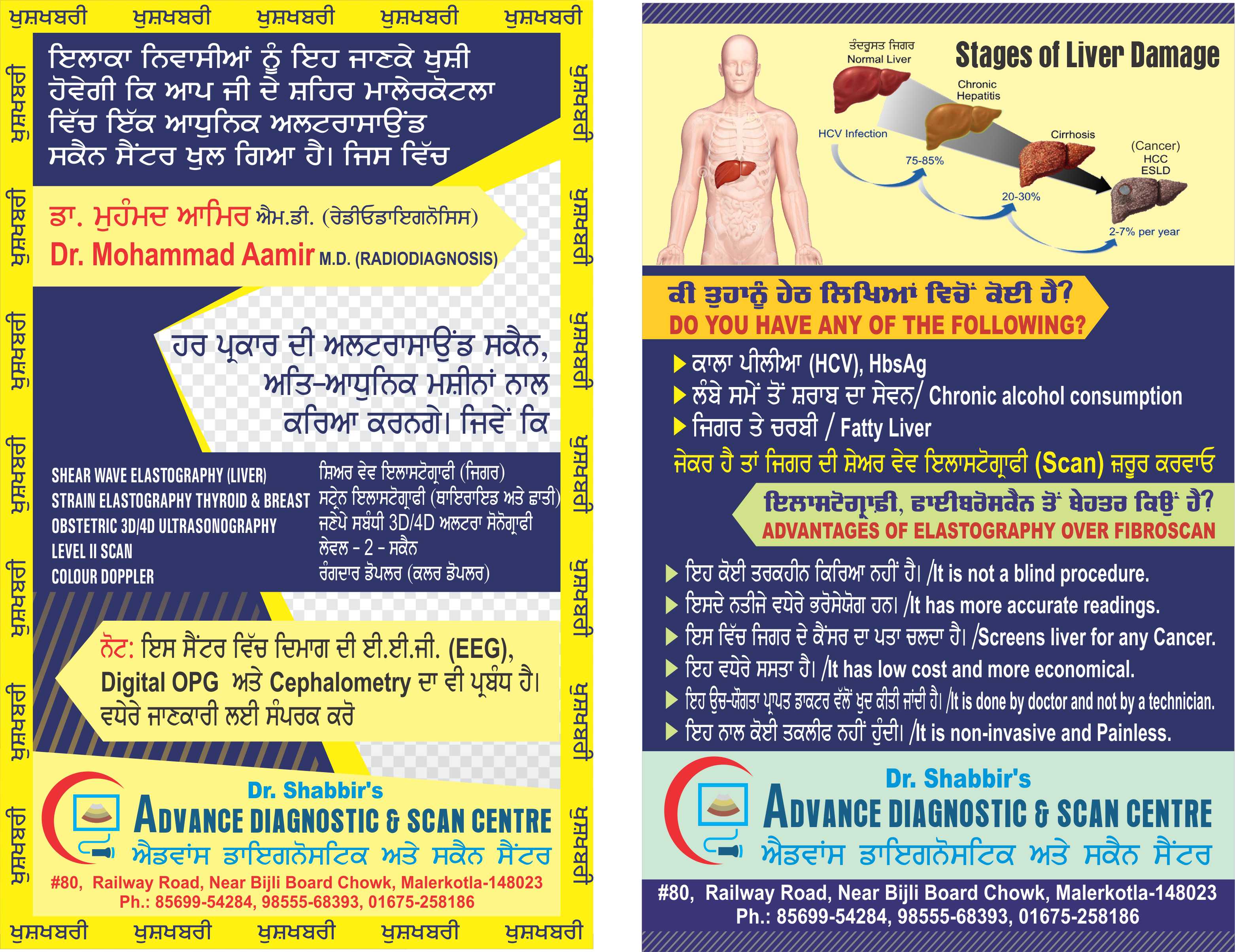
To Deliver 'the Best' Patient care with 'Passion and Empathy'
To be a caring Healthcare Trusted by Patients and Staff, Renowned for Clinical Excellence and Innovation
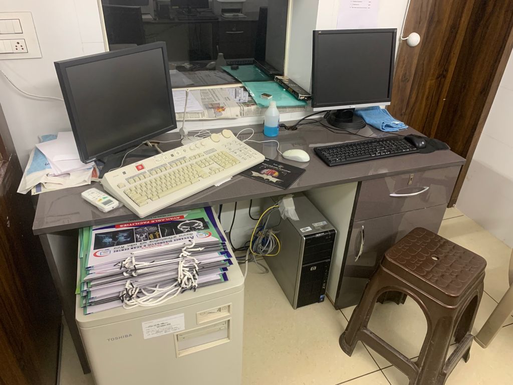
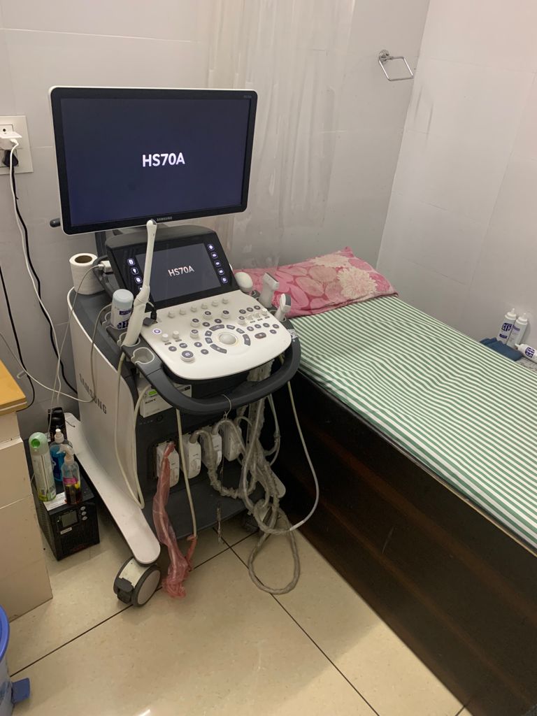
Online Cliniq provides the most advanced and versatile ultrasound diagnostics systems which can be used for imaging virtually every body part.
Read more
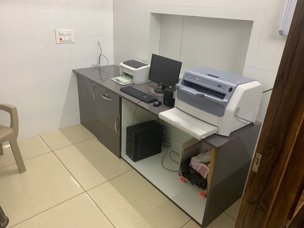
Online Cliniq provides the most advanced and versatile ultrasound diagnostics systems which can be used for imaging virtually every body part.
Read more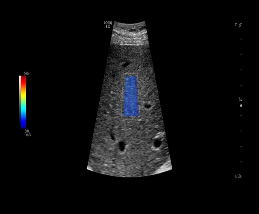
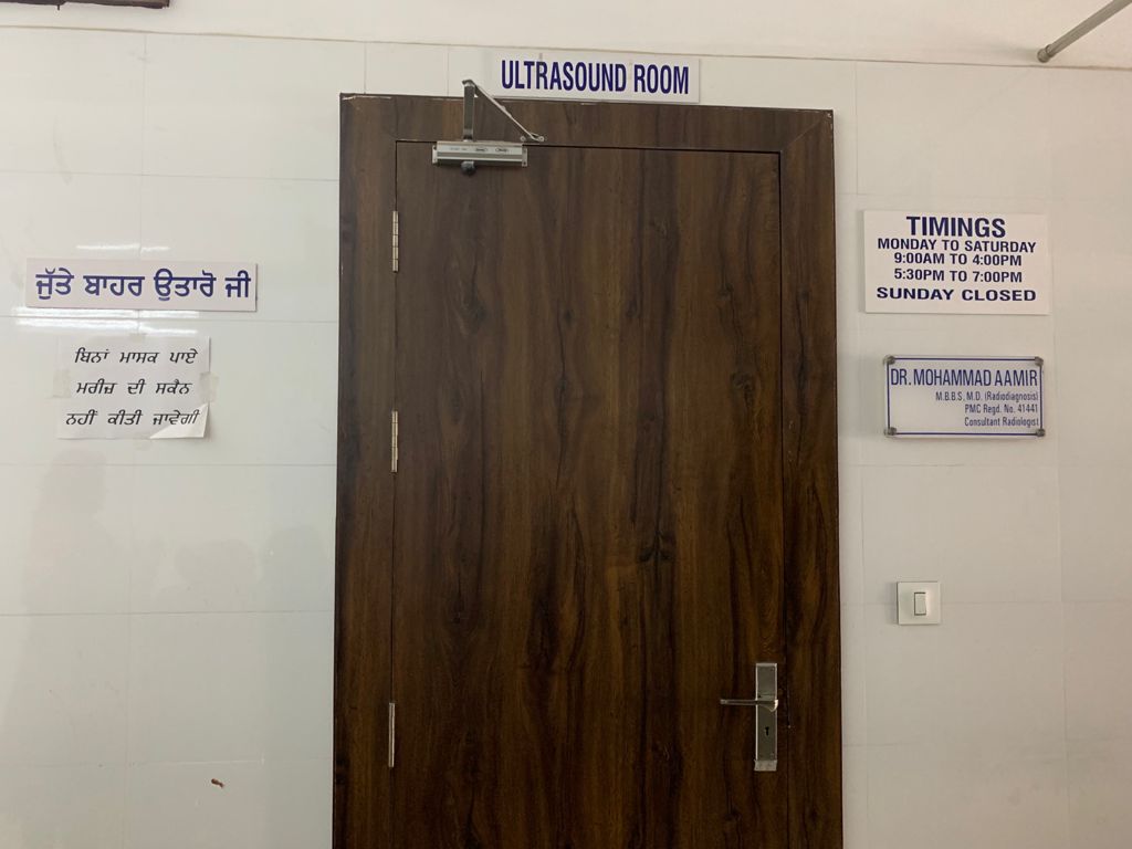
Online Cliniq provides the most advanced and versatile ultrasound diagnostics systems which can be used for imaging virtually every body part.
Read more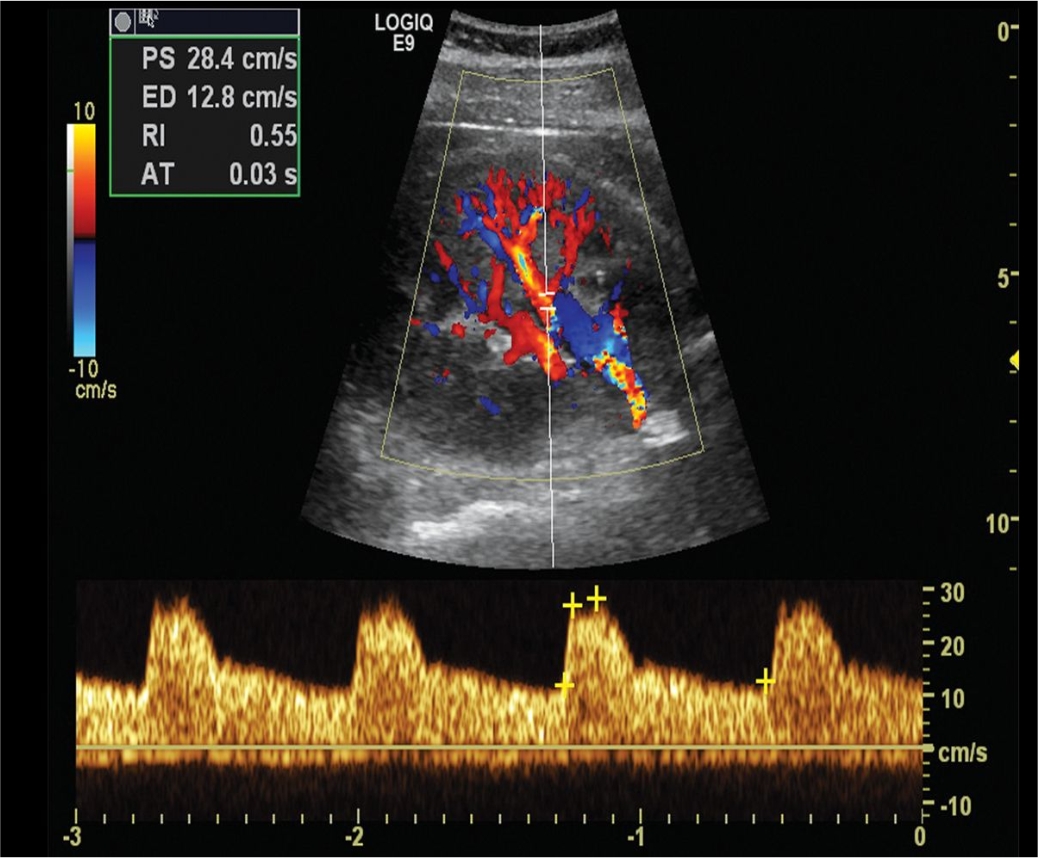
Online Cliniq provides the most advanced and versatile ultrasound diagnostics systems which can be used for imaging virtually every body part.
Read more
Online Cliniq provides the most advanced and versatile ultrasound diagnostics systems which can be used for imaging virtually every body part.
Read more
Online Cliniq provides the most advanced and versatile ultrasound diagnostics systems which can be used for imaging virtually every body part.
Read more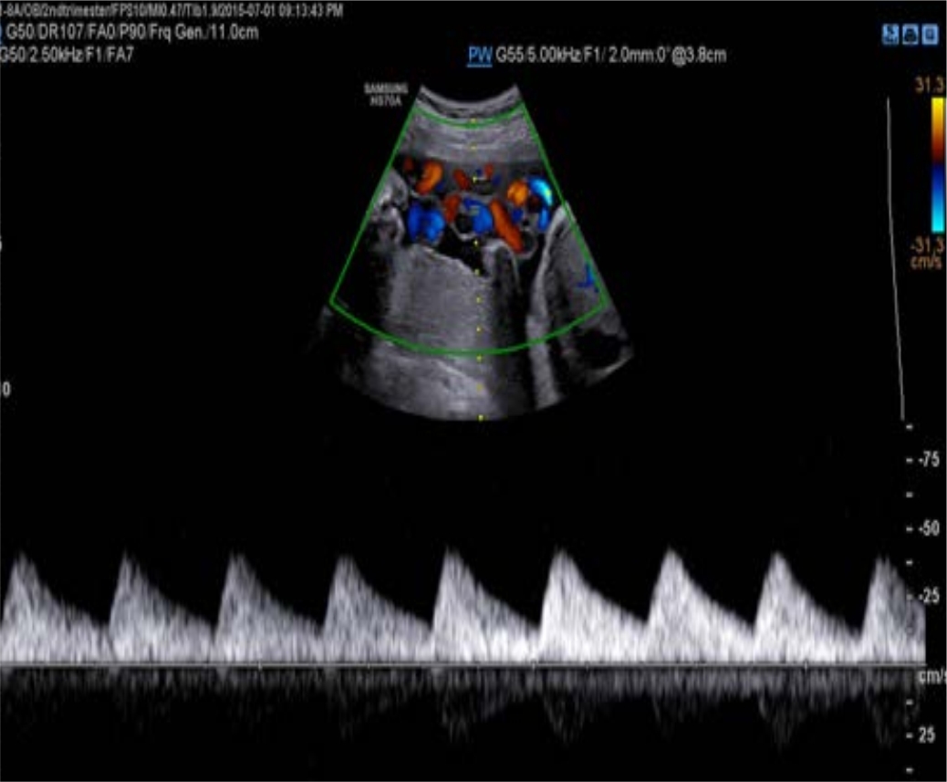
Online Cliniq provides the most advanced and versatile ultrasound diagnostics systems which can be used for imaging virtually every body part.
Read more
Online Cliniq provides the most advanced and versatile ultrasound diagnostics systems which can be used for imaging virtually every body part.
Read more


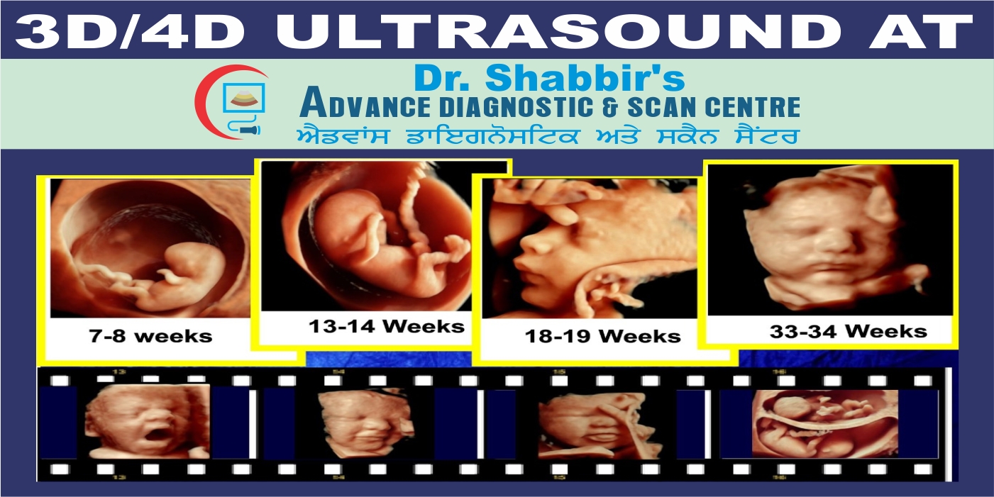
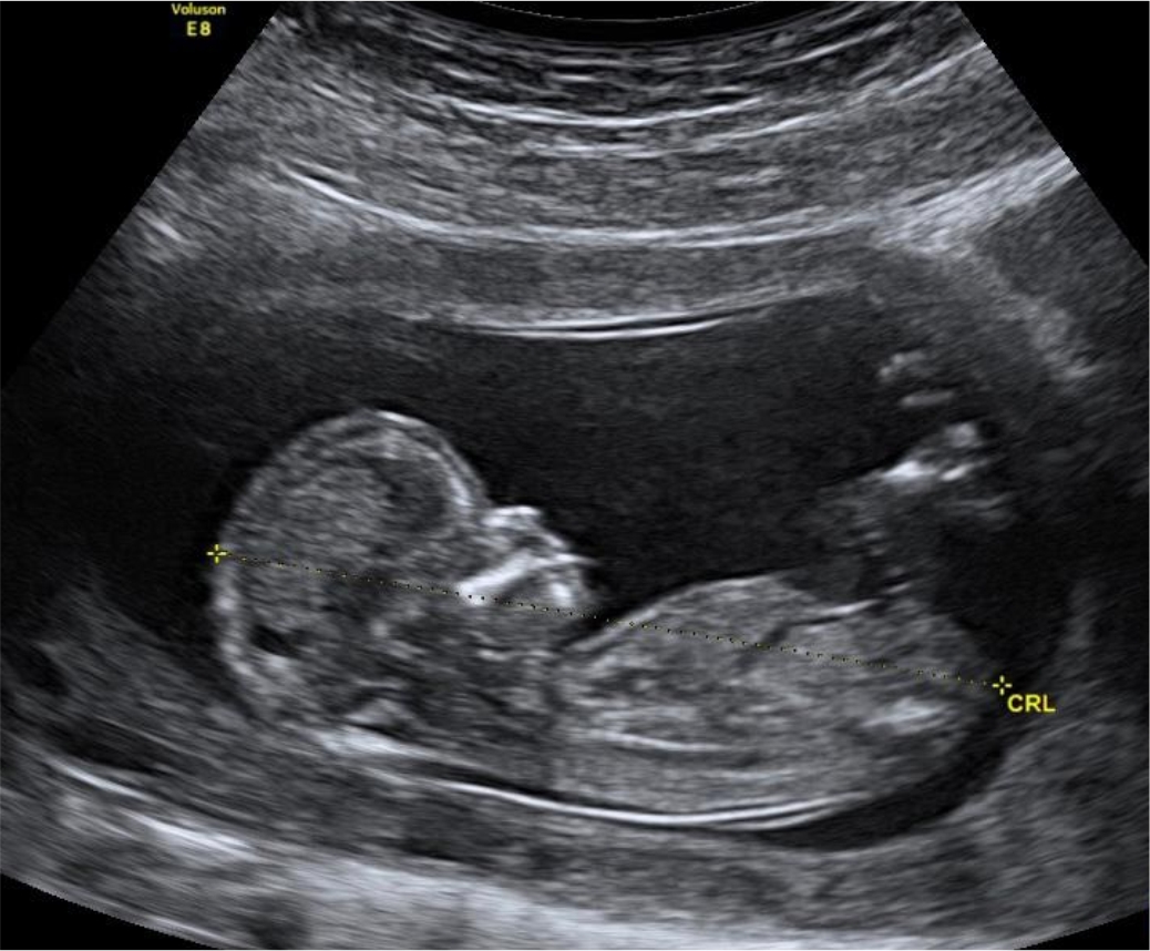
3D scans show still pictures of your baby in three dimensions. 4D scans show moving 3D images of your baby, with time being the fourth dimension. It's natural to be really excited by the prospect of your first scan. But some mums find the standard 2D scans disappointing when all they see is a grey, blurry outline. This is because the scan sees right through your baby, so the photos show her internal organs.
With 3D and 4D scans, you see your baby's skin rather than her insides. You may see the shape of your baby's mouth and nose, or be able to spot her yawning or sticking her tongue out.
3D and 4D scans are considered as safe as 2D scans, because the images are made up of sections of two-dimensional images converted into a picture. However, experts do not recommend having 3D or 4D scans purely for a souvenir photo or recording, because it means that you are exposing your baby to more ultrasound than is medically necessary. Some private ultrasounds can be as long as 45 minutes to an hour, which may be longer than recommended safety limits.
3D and 4D scans may nonetheless provide more information about a known abnormality. Because these scans can show more detail from different angles, they can help in the diagnosis of cleft lip. This can help doctors to plan the repair after birth.
3D scanning can also be useful to look at the heart and other internal organs. As a result, some fetal medicine units do use 3D scans, but only when they're medically necessary.
Most pelvic ultrasounds are performed using both the transabdominal and transvaginal approaches. Transabdominal ultrasound involves scanning through your lower abdomen. Transabdominal ultrasound usually provides an overview of the pelvis rather than detailed images. The transabdominal assessment is particularly helpful for the examination of large pelvic masses extending into the abdomen, which are not always well viewed with transvaginal ultrasound.
A small amount of ultrasound gel is put on the skin of the lower abdomen, with the ultrasound probe then scanning through this gel. The gel helps improve contact between the probe and your skin.
Transvaginal ultrasound is an internal ultrasound. It involves scanning with the ultrasound probe lying in the vagina. Transvaginal ultrasound usually produces better and clearer images of the female pelvic organs, because the ultrasound probe lies closer to these structures.
The transvaginal ultrasound probe is thin, about 2cm diameter. The probe is covered with a disposable protective sheath. A small amount of ultrasound gel is placed on the end of this probe. The probe is then gently inserted a short distance into the vagina. All transvaginal probes have been cleaned and sterilised according to recommended protocols.
Your privacy will always be respected during a pelvic ultrasound, especially the transvaginal examination. You will have a towel covering your lower body, in addition to wearing a gown during the transvaginal ultrasound.
You will always have a choice about whether transvaginal ultrasound is performed. If you have concerns about transvaginal ultrasound, please discuss this with your sonographer before your pelvic ultrasound begins.
We usually get better images during transabdominal ultrasound if the bladder is partially filled, so to help your examination we ask you to drink water prior to the assessment. Please empty your bladder 1 hour before your appointment, drink 2 glasses of water and try not to empty your bladder again until after your appointment. A full bladder moves bowel out from the pelvis into the abdomen, helping visualisation of the uterus and ovaries.
Your bladder should not be so full that it causes pain. If your bladder is very full and painful, you should empty a small amount so you are more comfortable. You will be able to empty your bladder after the transabdominal ultrasound is completed and before the transvaginal ultrasound begins.
Your doctor may find a pelvic ultrasound useful in the investigation of a number of problems including:
The pelvic ultrasound may not provide your doctor with all the answers to your problems, but it may be very helpful in diagnosis and management. Your referring doctor will decide if further treatment or tests are needed.
Assessment of fetal growth and well being in the third trimester, which routinely includes Doppler assessment of Middle Cerebral artery, Umbilical artery, Fetal venous return and maternal uterine arteries to assess any compromise in feto-placental and materno-placental circulations.
In general, Doppler scans look at the blood flow between you and your baby, to check whether he’s getting everything he needs to develop healthily
Uterine arteries are the vessels which carry blood to your womb (uterus). A uterine artery Doppler scan checks that enough blood is reaching your placenta.
Your baby needs plenty of nutrients and oxygen to grow at a healthy rate. Therefore, the walls of your uterine arteries should be stretchy, to allow as much blood through as possible. In pregnancy, these normally small arteries increase in size to allow more blood to reach your womb easily. This is called low resistance. If blood can’t get through to the placenta easily enough, your baby may not get the nutrients and oxygen he needs via the umbilical cord. Factors such as smoking, high blood pressure, hormone levels, and certain medications may increase the resistance in your artery walls.